 |
|
|
| |
| 05th May 2011 |
|
| 3D printing could become useful planning tool for neurosurgeons |
| |
| By Olivia Solon |
 |
| View Gallery |
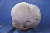
| 3D model of complex infant skull deformity (craniosynostosis) used for parent education and neurosurgical planning |
| |
| Advances in 3D printing have meant that radiologists can use the technique to quickly build affordable 3D models for neurosurgical planning. |
| |
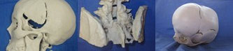
| Using ultra high-resolution CT scans, radiologists can transform the imagery into 3D solid models using a Z Corp 3D colour printer more commonly used in architecture and for rapid prototyping in engineering. |
| |
| The DICOM images (a standard file format for medical digital images) are converted to STL files readable by CAD software. This is then imported into Zprint software and printed by a Z Corp Spectrum Z510. |
| |
| The models help radiologists and surgeons identify defects that 2D images might not allow and give a clearer impression of the image. In addition to helping to plan procedures on complicated anatomy, it also helps physicians communicate with patients and their families. |
| |
| The technique is being employed at the Department of Radiology at Tripler Army Medical Centre in Hawaii in partnership with the Joint POW/MIA Accounting Command/Central Identification Laboratory. |
| |
| The Tripler doctors were previously sending data from Hawaii to mainland US to make models at vast expense and over a considerable time frame. |
| |
| Paediatric radiologist Lynne Reuss told Wired.co.uk: "Tripler neurosurgeons have found them very useful for complicated anatomy, but they have been used for less complicated anatomy as well, for example for planning prosthesis; in one case a model was used to make an appropriately sized cranioplasty flap (a replacement for a missing or damaged piece of skull)." |
| |
| Article gallery |
| View gallery |
 |
| |
| |